


Approximately 1% of the global population is affected by vitiligo, a condition characterized by the presence of white skin lesions. The majority, around 90%, of vitiligo cases fall into the nonsegmental category, which typically develops gradually in adulthood. Vitiligo involves the damage to melanocytes, specialized skin cells responsible for producing pigment. Melanocytes can be compromised anywhere in the body, affecting the functionality of organs in which they are found. Interestingly, melanocytes in both the uveal tract and the epidermis share an embryological origin, alongside physiological and anatomical similarities.
The authors of a recent study emphasize the systemic nature of vitiligo, highlighting its impact on ocular melanocytes within the retinal pigment epithelium and uveal tract, in addition to its effects on the skin.

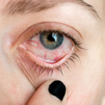
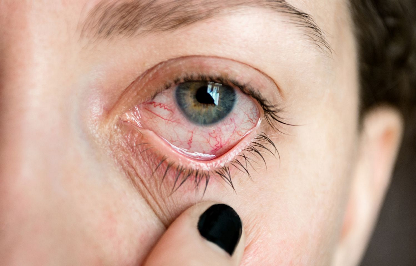
The study aimed to identify ocular changes in individuals with non-segmental vitiligo. The research findings align with earlier studies that have shown an increased likelihood of ocular abnormalities, uveitis (inflammation of the uvea), and subclinical inflammatory fundus depigmentation in people with vitiligo.
To conduct the study, researchers carried out a case-control investigation, enrolling 40 non-segmental vitiligo patients from outpatient clinics affiliated with Al-Azhar University in Egypt and 40 healthy controls. In the participant groups, males accounted for 52.5% of the vitiligo patients and 40% of the control group. Participants in both groups ranged in age from 18 to 40 years. Among the vitiligo patients, 55% had acro-facial lesions, while 45% had more widespread lesions. The mean duration of this idiopathic systemic autoimmune condition was 3.71 years among the study participants, ranging from 1 to 13 years. The investigation included both ophthalmic and dermatological assessments.

Key findings of the study include:
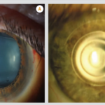
– 57.5% of vitiligo patients exhibited significant abnormalities during slit lamp fundus examinations, compared to only 6.3% of the controls. These abnormalities encompassed various issues such as a raised disc, tigroid fundus (fundus with streak-like patterns), conjunctival nevus (a benign growth on the conjunctiva), iris nevus (a benign iris tumor), and iris depigmentation.
– Central macular thickness, as determined by optical coherence tomography, displayed differences between the two groups, with the control group having the highest values. Refraction error values were also highest in the control group.

The authors stress the need for further research to explore potential risks and optimal treatment strategies for vitiligo and other dermatological conditions. In conclusion, the study found that although there were no notable differences in visual acuity, individuals with non-segmental vitiligo exhibited a significantly higher prevalence of ocular changes compared to the control group.